Week 6
The first week of July was a short week in terms of trapping. On Monday Ben, Izi, and I set up traps at the UMD Medical Campus Police Station, Queenstown Park, and the Patuxent Wildlife Refuge. The Patuxent Wildlife Refuge is the prettiest place we have set traps this trapping season by far! Now that it is July, we are going back through the 24 site locations to re-trap. As the summer progresses, the community distribution often changes resulting in different species of Culex peaking at different times. According to previous studies, Culex restuans peak first near the end of spring, while pipens peak in June and July.1
During the downtime of sorting and identifying Culex, Izi and I had the opportunity to learn more about other laboratory techniques of RNA isolation with an RNAeasy miniprep kit, along with qualifying controls used to determine the quantity of RNA isolated.
To start the RNA isolation process, β-Mercaptoethanol is mixed with Buffer RLT in a 2 ml flat-bottom tube with a ceramic bead. Then 30 mg of sample tissue is added and vortexed for homogenization. The next step is to lysate for three minutes through centrifugation. This generates a supernatant and a pellet of particulate tissue. With a micropipette, the supernatant is carefully collected without disturbing the pellet. I found this to be the hardest part, and it took me two attempts when I first tried. The supernatant is then divided into two angle-bottom tubes in 300 µl aliquots. Then 300 µl of 70% ethanol is added to both tubes and mixed by pipetting. The next part of RNA isolation involves a series of spin columns. The first spin column binds DNA to separate the RNA, while the RNAeasy spin column washes the RNA with various buffers before drying the RNA and adding RNAase-free water. Throughout the procedure, it was crucial to maintain aseptic technique and reduce the potential degradation of the RNA from RNAases which significantly reduce yield. Thus, I could not take pictures of the procedure to prevent contamination. Instead here are some pics that didn't make it into other blog posts!
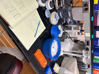
Here is a picture of the set-up for PCR 68. The diagram for what individual goes in each strip tube is written in my lab note book.
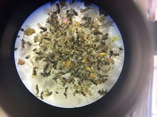
Here is a pile of mosquitos and other insects from one of the traps
where we got more than 200 mosquitos!


Comments
Post a Comment